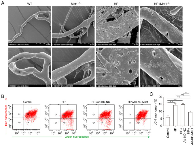
Figure 5.
Microvascular endothelial cell integrity and mitochondrial membrane potential of Ang II-treated endothelial cells. (A) Representative scanning electron microscopy images of mouse myocardial microvascular endothelial cells. (B) FACS measurement of mitochondrial membrane potential of CMECs. (C) Quantitative assessment of mitochondrial membrane potential of CMECs; n=3. *P<0.05 and **P<0.01. CMECs, cardiac microvascular endothelial cells; WT, wild-type; Mst1, mammalian ste20-like kinase 1; HP, hypertensive/hypertension; KD, knockdown; NC, negative control.

