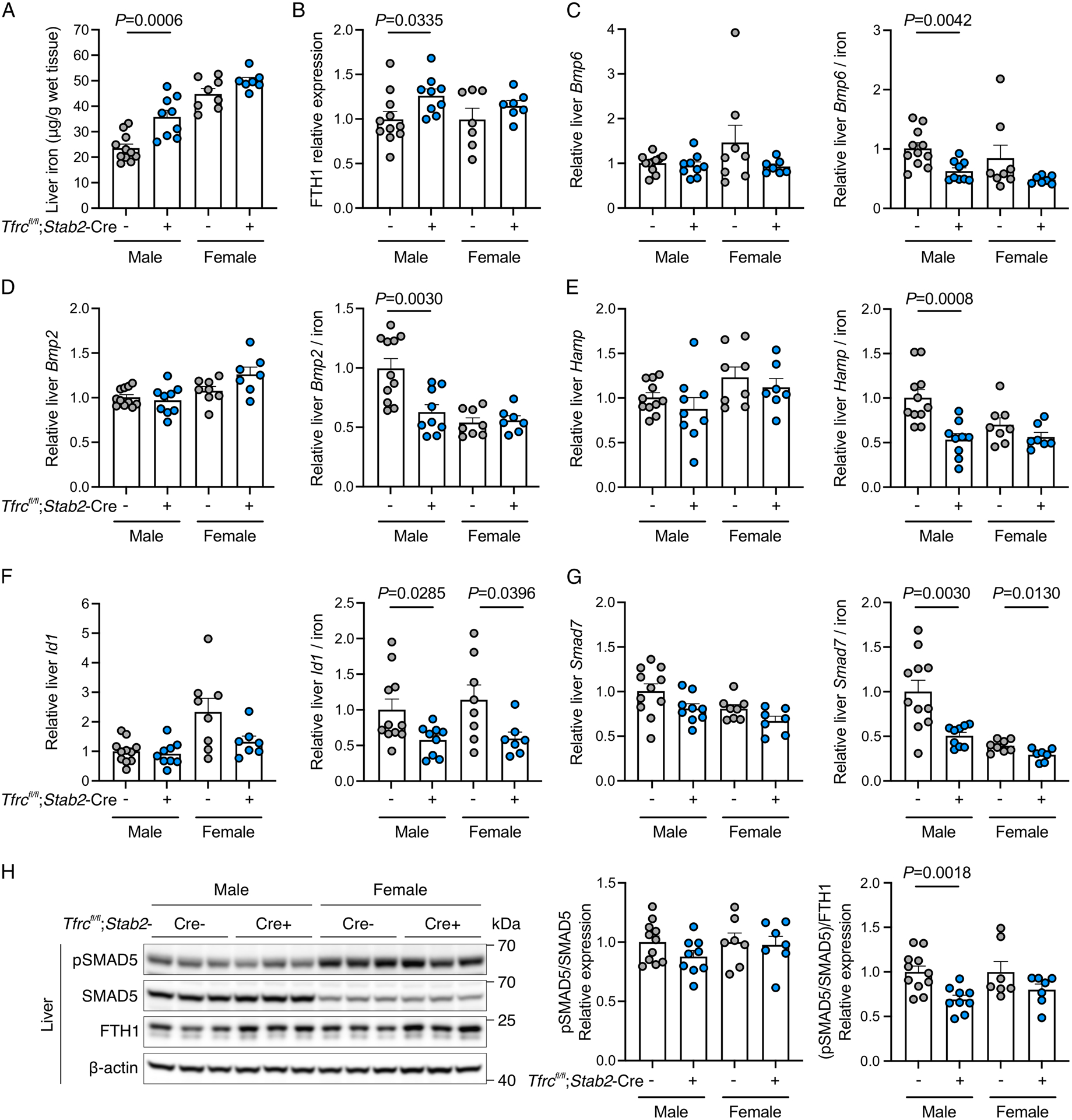
Figure 3. Functional role of endothelial TFR1 in iron sensing and hepcidin regulation in iron-limited conditions.

Three-week-old male (N=9–11/group) and female (N=7–8/group) Tfrcfl/fl;Stab2-Cre+ and littermate Cre− mice were fed a limited iron diet (20 ppm iron) for 9 weeks and analyzed at 12 weeks for: (A) liver iron concentration; (B) liver ferritin heavy chain (FTH1) relative to β-actin protein expression by Western blot and chemiluminescence quantitation; (C-G) hepatic Bmp6, Bmp2, Hamp, Id1, and Smad7 transcript levels relative to Rpl19 by qRT-PCR (left panels) and divided by liver iron concentration to control for iron-mediated regulation of these genes (right panels); (H) Western blot and quantitation of phosphorylated SMAD5 (pSMAD) relative to total SMAD5 (left panel) and divided by FTH1 levels to control for iron-mediated regulation (right panel). A representative blot is shown for the quantitation in panels (B) and (H). Graphs represent mean ± SEM with individual points indicating the number of animals per group. Statistical differences between sex-matched Tfrcfl/fl;Stab2-Cre+ and Cre- mice were determined by two-tailed Student’s t-test or Mann-Whitney U test for non-normally distributed values.
