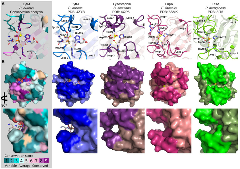
Figure 3.
Crystal structures of representative M23 metallopeptidases: LytM (blue; 4ZYB), Lysostaphin (violet; 4QPB), EnpA (magenta; 6SMK), and LasA (green, 3IT5). The M23 peptidases sequence conservation score was calculated in the ConSurf server and is displayed on LytM surface (PDB ID: 4ZYB; Landau et al., 2005). (A) Active sites and loop architectures are represented as cartoons. The active site residues are depicted as bold sticks and labelled accordingly. The zinc ion is shown as a yellow sphere. The LytM was crystallized in the presence of tetraglycine phosphinate, and the resulting complex served as a model for the substrate-binding mechanism. (B) The top view (upper panel) and site-zoomed view (bottom panel) of the corresponding surface represent the M23 structures.

