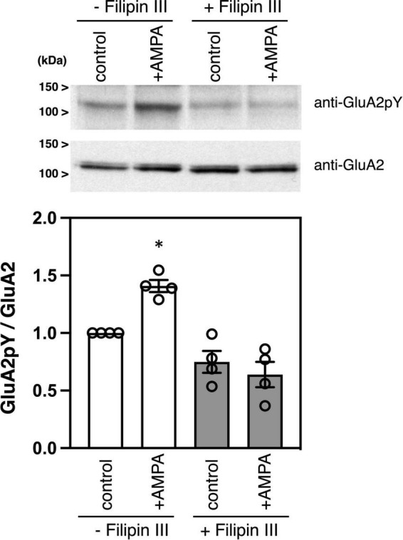
FIGURE 2.

Effects of filipin III treatment on AMPA-induced tyrosine phosphorylation of GluA2 in cultured cortical neurons. Cultured cortical neurons were treated with DMSO or 10 μg/ml Filipin III for 10 min, followed by stimulation with 100 μM AMPA for 10 min. Cell lysates from cultured cortical neurons were immunoblotted with anti-GluA2CpY or anti-GluA2 antibodies to quantify tyrosine phosphorylated GluA2 and GluA2 protein expression. Typical blots as representatives are shown (Top). The ratio of tyrosine phosphorylated GluA2 to GluA2 protein amounts is statistically analyzed (Bottom). Error bars represent SEM. *p < 0.05, compared with control.
