. 2022 Jun 29;28(15):3268–3276. doi: 10.1158/1078-0432.CCR-22-0666

Figure 3.

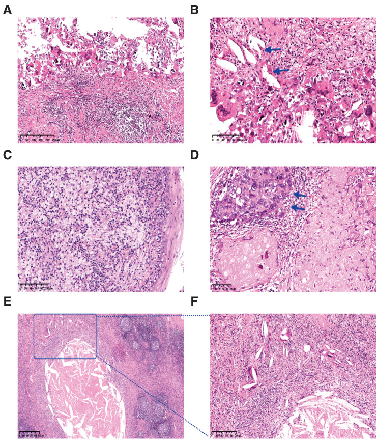
Figure 3. Significant tumor response was observed in the radical specimens. A and B, Fibrosis, lymphocyte infiltration, necrosis, cholesterol clefts (arrow), giant cell reaction, and calcification in primary tumor with pathological complete response (pCR). Hematoxylin–eosin. A, magnifications, ×100. B, magnifications, ×200. C, An aggregation of macrophages into the regression bed of HPV+ subject with pCR. Hematoxylin–eosin, magnifications, ×200. D, Acellular keratin and residual viable tumor (arrow) in the primary tumor with major pathological response (MPR). Hematoxylin–eosin, magnifications, ×200. E and F, Necrosis, histiocytes, cholesterol clefts, and giant cell reaction in metastatic lymph node with pCR. Hematoxylin–eosin. E, Magnifications, ×40. F, Magnifications, ×100. F, Is an enlargement within the square of (E).

Significant tumor response was observed in the radical specimens. A and B, Fibrosis, lymphocyte infiltration, necrosis, cholesterol clefts (arrow), giant cell reaction, and calcification in primary tumor with pathological complete response (pCR). Hematoxylin–eosin, magnifications, ×100 (A) and ×200 (B). C, An aggregation of macrophages into the regression bed of HPV+ subject with pCR. Hematoxylin–eosin, magnifications, ×200. D, Acellular keratin and residual viable tumor (arrow) in the primary tumor with major pathological response (MPR). Hematoxylin–eosin, magnifications, ×200. E and F, Necrosis, histiocytes, cholesterol clefts, and giant cell reaction in metastatic lymph node with pCR. Hematoxylin–eosin, magnifications, ×40 (E) and ×100 (F). F is an enlargement within the square of E.