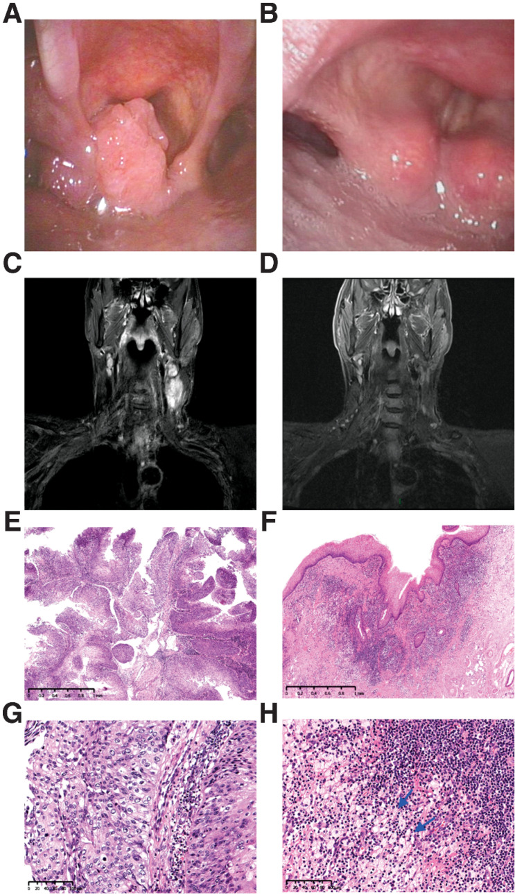
Figure 4.
Tumor regression in a 61-year-old man with stage IVA hypopharyngeal cancer (T2N2cM0, P21). The tumor was PD-L1 positive (combined positive score = 10) and originated in the left pyriform sinus. A, Pretreatment image showed a cT2 squamous cell carcinoma of the left pyriform sinus. B, Pretreatment MR imaging showed bilateral cervical lymph nodes. C and D, After three cycles of neoadjuvant chemoimmunotherapy, image at the time before surgery demonstrates near-complete clinical regression of primary lesion as well as cervical lymph nodes. E and F, Pathological findings of biopsy specimens of hypopharyngeal masses before treatment. The tumor is non-keratinizing squamous cell carcinoma with papillary features. Hematoxylin–eosin, magnifications, ×30 (E) and ×200 (F). G and H, Histopathologic images of the resection specimen after treatment. Fibrosis, lymphocyte infiltration, and histiocytes aggregation (arrow) were found in the regression bed, and no cancer residue was found (pathological complete response).Hematoxylin–eosin, magnifications, ×30 (G) and ×200 (H).

