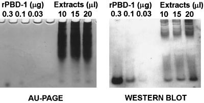
FIG. 3.
Determination of PBD-1 concentration in pig tongue epithelia. (Left panel) Porcine tongue epithelial extracts (10 to 20 μl) were subjected to AU-PAGE and stained with Coomassie blue. Recombinant PBD-142 (0.03 to 0.3 μg) was used as a standard. (Right panel) Proteins on an AU-PAGE with the same loading pattern were blotted to an Immobilon-P membrane; the blots were probed with rabbit anti-PBD-1 antibody and developed as described in Materials and Methods. Recombinant PBD-142 (0.03 to 0.3 μg) was used as a standard for the semiquantitative analysis of PBD-1 concentration.

