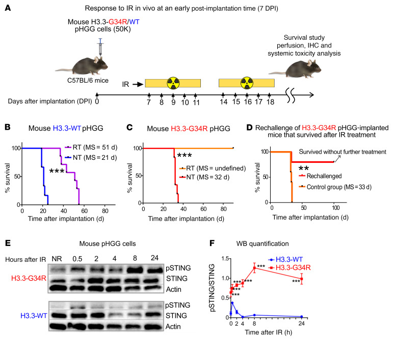
Figure 10. H3.3G34R pHGG shows an improved therapeutic response to RT, and DNA damage in these cells mediates cGAS/STING pathway activation.
(A) H3.3-G34R and H3.3-WT mouse cells were implanted to generate allogenic pHGG in mice. Mice were subjected to 20 Gy RT starting on day 7 after implantation according to the schedule indicated in the scheme (2 Gy/d, 10 days). (B) Kaplan-Meier survival plot of H3.3-WT–bearing mice treated with RT compared with NT mice. (C) Kaplan-Meier survival plot of H3.3-G34R–bearing mice treated with RT compared with NT mice. (D) Kaplan-Meier survival plot of H3.3-G34R–bearing mice that survived after RT treatment as indicated in C and that were rechallenged by implantation of H3.3-G34R cells into the contralateral hemisphere. (E) STING (phospho-Ser365) levels in H3.3-G34R and H3.3-WT mouse cells at different time points after 3 Gy IR. (F) Quantification of the Western blot (WB) results represented in E. **P < 0.01 and ***P < 0.005; analysis of MS from the Kaplan-Meier curve; n = 5 mice/group (B–D). Data were analyzed by log-rank (Mantel-Cox) test (B–D) and unpaired t test (F). Data in F represent the mean ± SD of 3 technical replicates.

