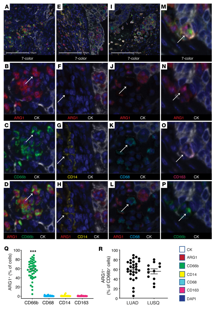
Figure 1. Arginase 1 is predominantly located within neutrophil lineage cells in human NSCLC.
(A–P) Representative images from NSCLC cases (n = 44) stained for CD66b (green), CD68 (cyan), ARG1 (red), CD14 (yellow), CD163 (pink), AE1/AE3 (CK, white), and with DAPI (blue). Stained slides were imaged on the Vectra 3.0 platform and analyzed using HALO. (A–D) Depict ARG1 positivity within the CD66b+ population. (E–H) Depict ARG1 negativity within the CD14+ population. (I–L) Depict ARG1 negativity within the CD68+ population. (M–P) Depict a macrophage triple-positive for ARG1, CD163, and CD66b. Original magnification, ×10 (A, E, and I) and ×40 (all other panels). Scale bars: 100 μm. (Q) Percentage of ARG1+ cells in CD66b+ (green), CD68+ (cyan), CD14+ (yellow), and CD163+ (pink) cells quantified from FFPE human NSCLC slides, n = 44. ***P < 0.001 by 1-way ANOVA with Tukey’s post hoc test. (R) Percentage of ARG1+ cells in the CD66b+ population for LUAD (n = 32) and LUSQ (n = 12).

