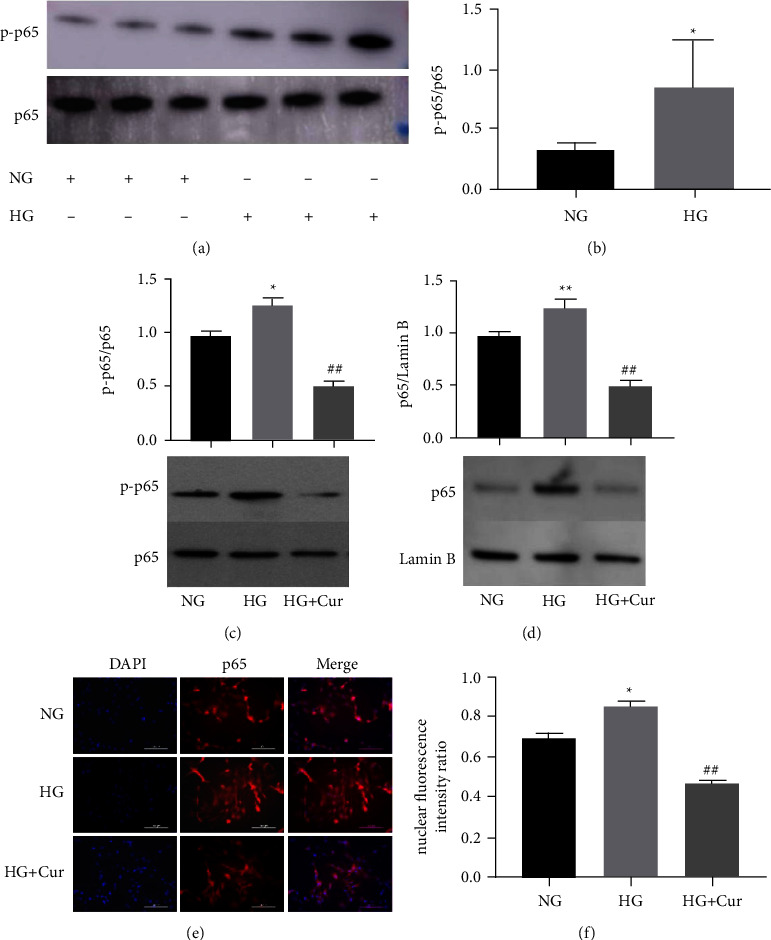
Figure 4.

Cur regulated NF-κB signaling in BMSCs. (a)–(b) Western blot of p65 and p-p65 in BMSCs treated with NG and HG. (c) Western blot of p65 and p-p65 in BMSCs treated with NG, HG, and HG + Cur. (d) The nuclear protein levels of p65 were detected by western blot in NG, HG, and HG + Cur group. (e) BMSCs were fixed and incubated with an anti-p65 antibody. Nuclei were stained by DAPI. The nuclear and cytoplasm images were merged in the same visual field. (f) Quantitative analysis of the nuclear translocation of p65. Data are presented as the mean ± SD from at least three independent experiments. ∗p < 0.05 and ∗∗p < 0.01 versus NG group. #p < 0.05 and ##p < 0.01 versus HG group.
