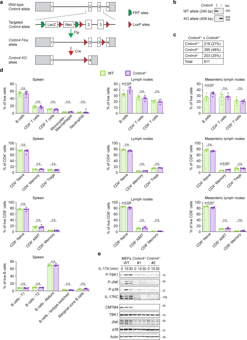
Extended Data Fig. 7. Characterization of CMTM4-deficient mouse.
a, Schematic representation of Cmtm4 knockout strategy. Cmtm4 coding exons and position of FRT and LoxP sites are indicated. b, Example of routine PCR genotyping of wild-type and Cmtm4−/− mice. c, Number of pups with the indicated genotype born to Cmtm4+/− parents. d, Flow cytometry analysis of different immune subtypes in indicated immune organs isolated from 8-12 weeks old Cmtm4+/+ (WT) or Cmtm4−/− mice. B cells (CD19+) were divided in T1 (IgM+, CD23−, CD1d−), T2 (IgM+, CD23+, CD1d−), marginal zone B cells (IgM+, CD23−, CD1d+), mature (IgM−, IgD+), and isotype switched (IgM−, IgD−) cells. Myeloid cells (CD3−, CD19−, NK1.1−, CD11b+) were divided into neutrophils (CD11c−, Ly6G+) and monocytes - macrophages (CD11c−, Ly6G−). CD8+ T cells were divided in naive (CD44−), memory (CD44+, CD49d+), and antigen-inexperienced memory T (AIMT) (CD44+, CD49d−) cells. CD4+ T cells were divided in Tregs (FoxP3+), naive (FoxP3−, CD44−, GITR−) and memory (FoxP3−, CD44+, GITR+) cells. n = 6 mice per group in three independent experiments. Mean + s.e.m., two-tailed Mann–Whitney test. n.s., not significant. e, Immunoblotting analysis of lysates of mouse embryonic fibroblasts (MEFs) isolated from Cmtm4+/+ or Cmtm4−/− sibling embryos stimulated with SF-IL-17A (500 ng ml−1) for indicated time points. Data (e) are representative of two independent experiments.

