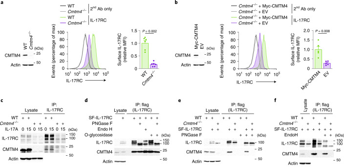
Fig. 2. CMTM4 mediates IL-17RC glycosylation and plasma membrane localization.
a,b, Immunoblotting analysis of CMTM4 and actin and flow cytometry analysis of IL-17RC surface expression in wild-type and Cmtm4−/− ST2 cells (a) or Cmtm4−/− ST2 cells transduced with expression vectors coding for Myc-CMTM4 or EV (b). c, Immunoblotting analysis of samples isolated via IP with an IL-17RC antibody from lysates of wild-type and Cmtm4−/− ST2 cells stimulated with IL-17A (500 ng ml–1) for 15 min or left unstimulated. d–f, Immunoblotting analysis of samples isolated through Flag IP from lysates of wild-type and Cmtm4−/− ST2 cells expressing SF-IL-17RC that were treated with O-glycosidase (d), PNGase F (d, e) or Endo H (d, f) as indicated. Data are representative of two (c–f) independent experiments. Flow cytometry data are represented as mean + s.e.m. from six (a) or five (b) independent experiments. Two-tailed Mann–Whitney test. MFI, median fluorescence intensity.

