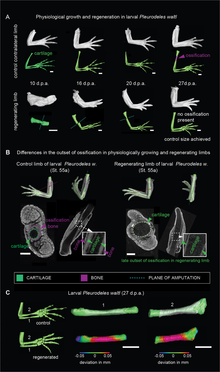
Fig. 2. The onset of ossification in normally developing limbs differs from regeneration.
A Micro-CT scans and 3D models of regenerating limbs in larval Pleurodeles waltl. Fully patterned larval limbs (stages 52a to 54) were amputated unilaterally, and limb regeneration was assessed at 10, 16, 20 and 27 days post-amputation (d.p.a.). The top panel shows physiological growth corresponding to contralateral control limbs. Note the ossification occurred in the control limbs while the regenerating limbs remained cartilaginous. Cyan dotted line point at the amputation plane. Scale bars, 500 µm. B Micro-CT scans and segmented 3D models with depicted slice planes showing a delayed outset of ossification in regenerating larval limb of Pleurodeles waltl. A representative contralateral control (left) and a regenerating limb (right) show the respective presence and absence of ossification in the humerus. Note the presence of chondrocytes underneath the ossified layer in the control limb. Green color represents the cartilage, and magenta color represents the bone. White dotted line marks the area that is magnified in the insets. Scale bars, 200 µm. C 3D comparisons of shapes of normally developed and regenerated skeletal elements from the forelimb of larval Pleurodeles waltl. Note that regenerated skeletal parts have increased diameter, the shape differences are presented as a heat-map of shape deviation. Scale bars, 500 µm.

