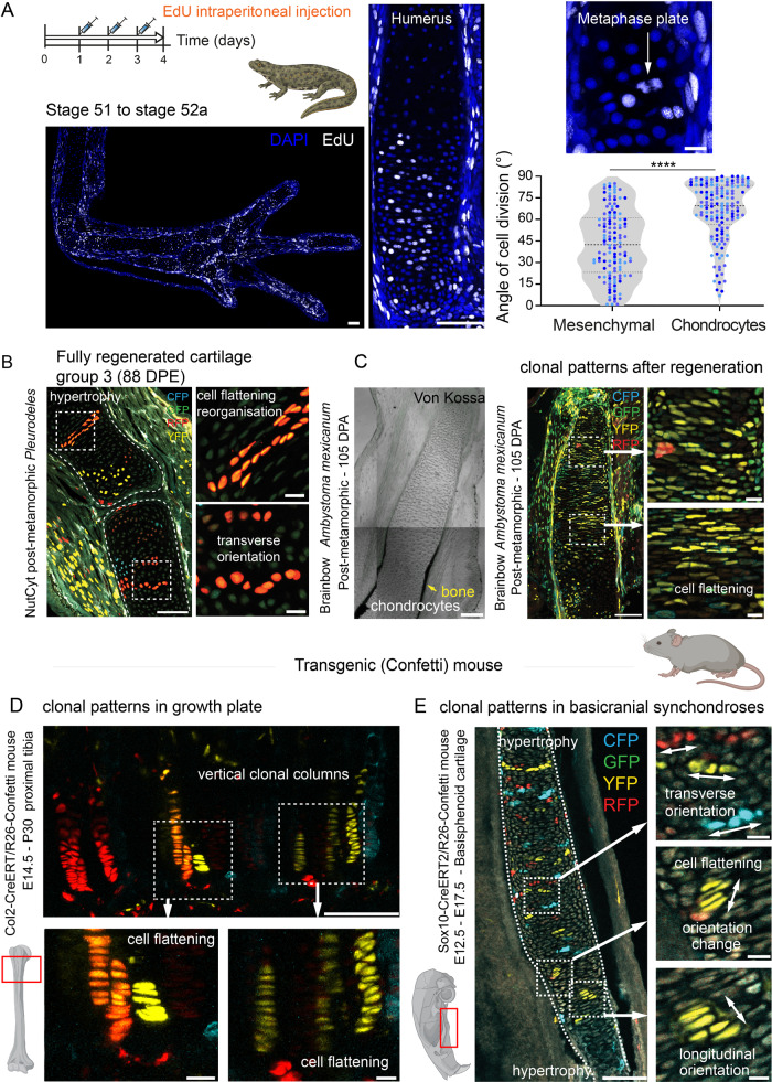
Fig. 4. Orientations of cell divisions and chondrogenic clones during salamander limb development and regeneration resemble the mammalian basicranial synchondroses.
A EdU pulse-chase performed at several time points (stage 51 to 52a is shown) in physiologically growing larval limbs of Pleurodeles waltl showed that the vast majority of chondrocyte cell divisions were oriented transversally. EdU-labelled doublets indicate cell division (shown in the right panel). Scale bar, 200 µm(left); 50 µm(center); 25 µm(right). The violin plots display differences in cell division/repositioning of cells found in rod-shaped skeletal elements of all species and processes analysed (see Fig. 2A, S5H, S6E). Two tailed t-test, ****p < 0.0001. Each data point represents orientation of a cell division, measured from n = 3 different limbs; median and quartiles are represented as dashed and dotted lines, respectively. B Transverse orientation of clonal chondrocytes in regenerating long bones of post-metamorphic Pleurodeles. Cell flattening at ossification onset correlates with absence of longitudinally oriented clones typical for mammalian long bones (see Fig. S6A). C Von Kossa staining of bone tissue in the regenerating limb of an experimentally-induced post-metamorphic Ambystoma mexicanum at 105 d.p.a. Yellow arrow points at mineralized layer with chondrocytes underneath. Next to it (right), neighboring section shows traced chondrocytes. D Proximal tibial growth plate is shown with the resting stem cell zone at the lower edge of the image. The recombination was induced at E14.5 in Col2CreERT2/R26Confetti embryos, when the limb is patterned, and the skeletal elements are made of stratified cartilage. The clones were analyzed at P30. A dotted line marks areas from magnified insets. Note the longitudinally-oriented chondrocytic clones containing proliferative flattened cells near hypertrophic zone. E Lineage tracing in mouse basicranial synchondroses highlights clonal arrangements. The recombination was induced at E12.5 in Sox10CreERT2/R26Confetti embryos, and analyzed at E17.5. The basicranial cartilage undergoes ossification at E17.5 and allows observing cell dynamics in synchondroses. A dotted white line marks areas in magnified insets. Note the presence of transversally-oriented chondrocytic clones within basisphenoid, and cell flattening and repositioning near hypertrophic zone. The patterns in D and E were observed independently in 10 or more individual embryos from three litters. Scale bars are 100 µm, in small square magnified insets the bars are 10 µm.

