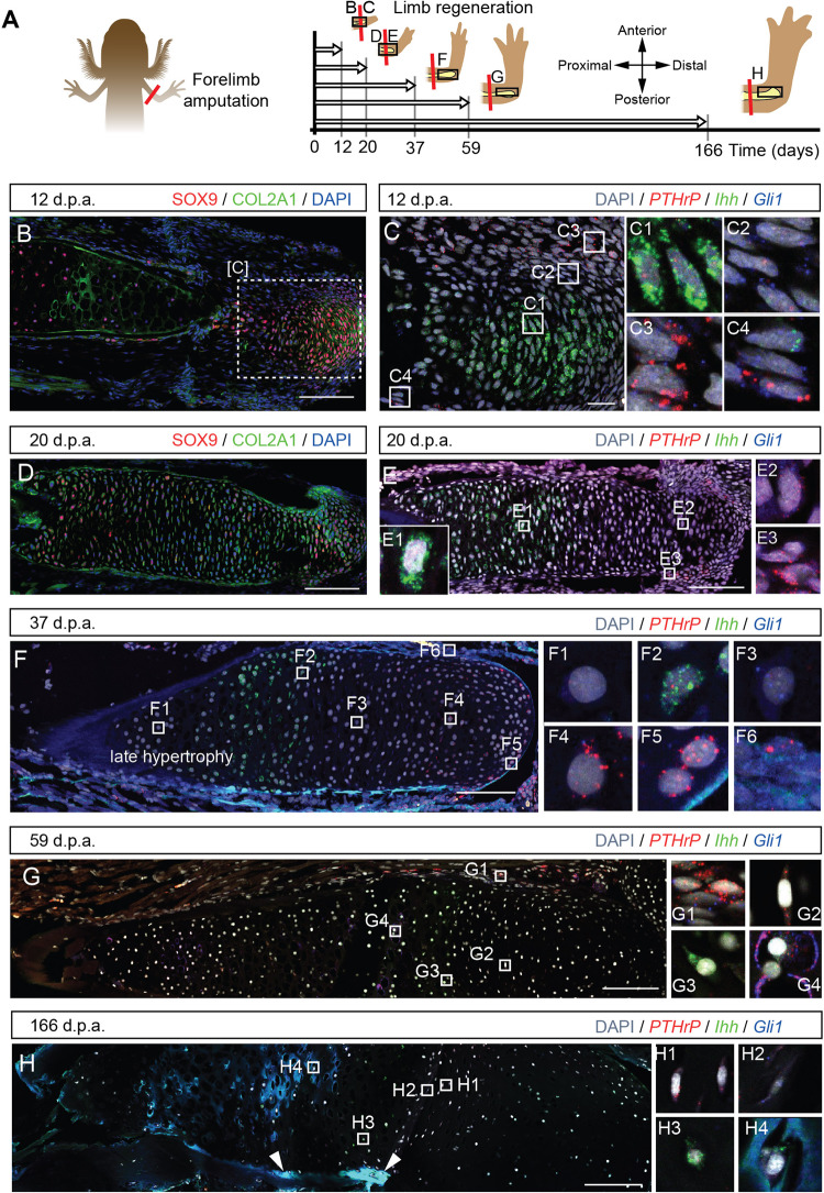
Fig. 6. Dynamic expression of the PTHrP-Ihh loop components during skeletal regeneration.
A Experimental outline for assessment of the PTHrP-Ihh loop during regeneration in Pleurodeles waltl. Larvae at stage 54 underwent unilateral amputations. The regenerating limbs and contralateral controls were collected at the selected time points. Note that the axis drawn in A applies to all the pictures of the regenerating humerus in the figure (B–H). B At 12 days post-amputation (d.p.a.), the core of the blastema showed SOX9+/Col2A1+ emerging chondrocytes. C At 12 d.p.a., a central group of Ihh+ (C1) is wrapped by a layer of Gli1+ (C2) cells followed by a layer of PTHrP+ cells (C3). This pattern was also observed in periskeletal cells surrounding the stump bone (C4). D At 20 d.p.a., the expanding humerus wrapped the stump bone and consisted of SOX9+/Col2A1+ chondrocytes. E At 20 d.p.a., Ihh+ pre-hypertrophic chondrocytes occupy a wide region proximal to the amputation plane (E1), while double-labelled Gli1+/PTHrP+ cells were found both in the distal portion of the humerus (E2) and in the perichondrium (E3). F At 37 d.p.a., we detected the first chondrocytes devoid of PTHrP-Gli1-Ihh (F1). Ihh+ expression was maintained in the pre-hypertrophic chondrocytes (F2), followed by Gli1+ cells (F3). PTHrP+ was strongly detected in periarticular chondrocytes in the epiphysis (F4, F5). Perichondrial cells expressed PTHrP and Gli1 at this stage (F6). G At 59 d.p.a., PTHrP and Gli1 perichondrial cells were still present (G1). The PTHrP+ expression was reduced in the epiphysis (G2), and the Ihh+ expression domain occupied most of the humerus (G3). We observed the first hypertrophic chondrocytes in the regenerates containing PTHrP and Gli1 puncta (G4). H At 166 d.p.a., the expression patterns of Ihh, PTHrP and Gli1 in the cartilage resembled those found in the contralateral control limbs. The PTHrP+ expression was restricted to fewer cells (H1), followed by scarce Gli1+ cells (H2) and further the Ihh+ cells (H3). We observed multiple hypertrophic chondrocytes in patches of the regenerate containing PTHrP and Gli1 puncta (H4). Arrowheads point to ossification occurring in patches. Scale bars: 200 µm (B, D–H) and 50 µm (C).

