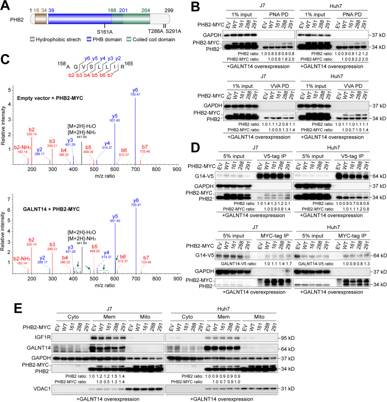
Fig. 4. Serine residue 161 of PHB2 serves as a target site for GALNT14-mediated O-glycosylation.
A Schematic representation of predicted PHB2 domains and residues as O-glycosylation hotspots. B PNA- or VVA-mediated pulldown of PHB2 was performed using lysates of cells transfected with the indicated plasmids. C Spectrum of Serine-161-containing peptides derived from LC/MS/MS. Green bars and arrows indicate that there were certain uncharacterized modifications on the fragmented peptides. m/z, the mass-to-charge ratio, where m is the molecular or atomic mass number and z is the charge number of the ion. D Western blot analysis following co-immunoprecipitation using samples of HCC cells transfected with the indicated plasmids. E Western blot analysis of subcellular fractions fractionated from cells transfected with the indicated plasmids, including cytosolic (Cyto), membranous (Mem) and mitochondrial (Mito). IGF1R was used as a specific marker for the membrane fraction, while VDAC1 was used as a marker for the mitochondrial fraction.

