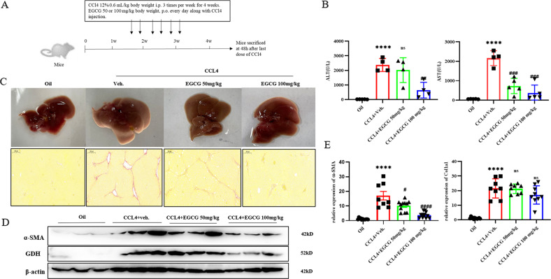
Fig. 3. Glutaminolysis activates myofibroblastic HSCs in chronically injured fibrotic livers in mice.
A Animal experimental procedures. B ALT and AST levels in serum. C Representative pictures of liver morphology in each group at sacrifice and Sirius red staining (scale bars = 200 μm); red indicates fibrosis. D Western blot analysis of the protein expression of α‐SMA, Col1a1, GDH and SIRT4. E Real‐time quantitative PCR analysis of the mRNA expression of fibrotic genes. The data are reported as the mean ± SD. *p < 0.05; **p < 0.01; ***p < 0.001; ****p < 0.0001 versus the oil group. #P < 0.05, ##P < 0.01, and ###P < 0.001 versus the CCL4 + Veh group.

