Figure 1.
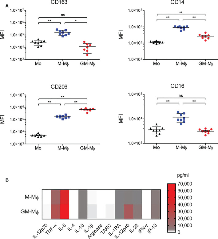
CD163, CD14, CD206 and CD16 analysis by flow cytometry during macrophage differentiation in GM-CSF or M-CSF. Human monocytes were cultured with 60 ng/ml M-CSF or GM-CSF for 6 days. (A) CD163, CD14, CD206 and CD16 expression was assessed by flow cytometry on day 0 in monocytes (MO) and day 6 in M-Mϕ and GM-Mϕ. Data are mean fluorescence intensity (MFI) from eight independent experiments (mean ± SD, N=8). (B) Results of flow cytometry of cytokine secretion in M-Mϕ and GM-Mϕ after lipopolysaccharide stimulation represented as a heatmap (mean ± SD, N=9). Mo, monocytes; M-Mϕ, M-CSF–induced macrophages; GM-Mϕ, GM-CSF–induced macrophages. ns = not significant, *p ≤ 0.05 and **p ≤ 0.01.

