Figure 1.
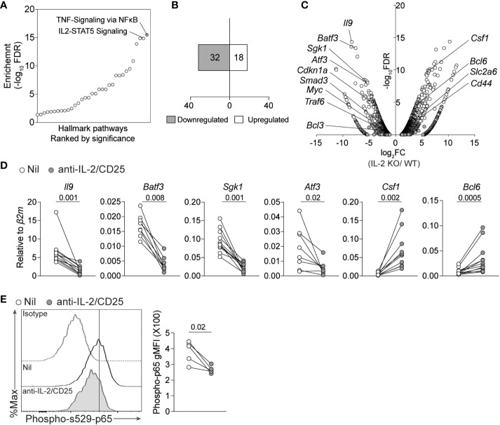
IL-2 deficiency results in a reduced NF-kB-associated gene signature in Th9 cells. (A) Pathway analysis from bulk-RNA-seq analysis of Th9 cells polarized from WT or IL-2-KO mice previously published (GSE41317). (B) Number of up- and down- regulated NF-kB signature genes (Fold change >2, p-value<0.05). (C) Volcano plot depicting selected genes regulated by NF-kB that are differentially expressed in IL-2 KO vs WT Th9 cells. (D) Th9 cells were cultured in standard or IL-2-limiting conditions, and 4 days after initial activation total RNA was isolated for qPCR validation of selected NF-kB-associated genes obtained from panel (A) Data in panel (D) are representative of naive T cell cultures from 8-11 individual mice pooled from 3 individual experiments. (E). Representative histograms and quantified geometric Mean Fluorescence Intensity (gMFI) of phospho- s529-p65 staining in cells cultured as in (D) Each data point represents values obtained from T cells isolated from one mouse, for a total of n=5 mice. Data were considered significantly different when paired t-test p value was ≤0.05.

