Figure 5.
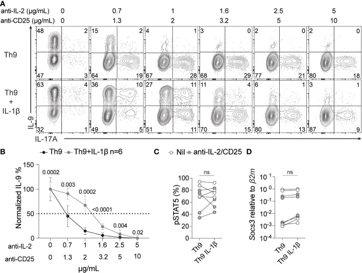
IL-1β signaling enhances IL-2 sensitivity during Th9 differentiation. Th9 cells were cultured in the presence or absence of IL-1β and increasing amounts of anti-IL-2/CD25 and IL-9 and IL-17A were measured by ICS at day 5 of culture. (A) Representative contour plots of intracellular IL-9 and IL-17A after restimulation with PMA and ionomycin. (B) Quantification of %IL-9+ Th cells at day 5 of culture from panel A, p<0.05 by paired Student’s t-test was considered significant, error bars represent SD. pSTAT5 protein (C) and Socs3 mRNA (D) levels of Th9 cells with or without IL-1β at no IL-2/CD25 blockade (Nil) or at 1μg/ml anti-IL-2/2μg/ml anti-CD25 (anti-IL-2/CD25). Data in panel A and B are representative of naïve T cell cultures from 6 mice and pSTAT5 and Socs3 data are representative of cultures from 4-6 mice from 2 pooled experiments. ns, non-significant.

