
FIGURE 2.
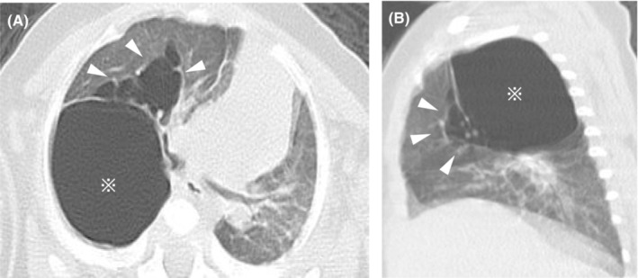
Chest computed tomography performed at 5 months of age demonstrates a large 30 × 25 mm monocystic space with a clear rim (asterisk) and multiple cystic air spaces with a line‐and‐dot pattern (white arrowheads) in the upper right lobe (A, B).