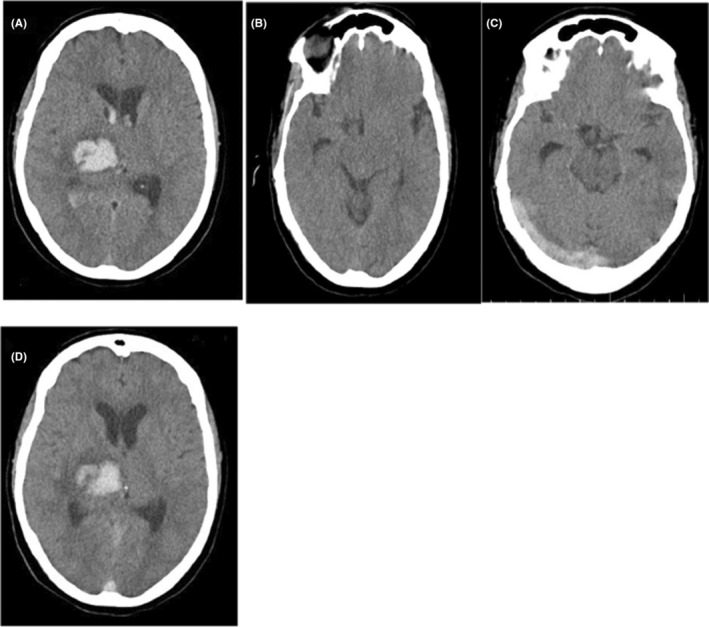
FIGURE 1.

(A) Brain computed tomography (CT) scan at the time of admission demonstrating intra cranial hemorrhage at the right thalamus and basal ganglia with expansion to the lateral ventricles. (B) at the level of the transverse and sigmoid sinus, there is no evidence on behalf of CVST. (C) On the seventh day after admission, a brain CT scan showed the cord sign and (D) a good absorption process for his ICH.
