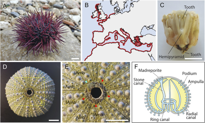
FIGURE 1.
General information on Paracentrotus lividus adults. (A) Photography of an adult specimen of the sea urchin species P. lividus. In the wild, P. lividus adults can be purple (as shown here), green, or brown. (B) Schematic representation of the geographical distribution of P. lividus, based on the Ocean Biodiversity Information System (OBIS, 2021). (C) Photography of a dissected masticatory apparatus of a P. lividus adult (i.e., Aristotle’s lantern). (D) Photography of the calcitic endoskeleton of a P. lividus adult. No matter the outer color of the adult, its endoskeleton is always green, more or less pale. (E) Close-up of the aboral surface and the central disk of the calcitic endoskeleton of a P. lividus adult, corresponding to the region highlighted by the yellow box in (D). In (E), note that the endoskeleton is composed of five ambulacra separated from each other by five interambulacra. Note further that each of the five gonopores (pores through which the gametes are released) is held by a genital plate (marked by the cyan and red arrowheads). One of the five genital plates is bigger than the others, it is the madreporite (marked by the cyan arrowhead), which corresponds to a sieve plate enabling water to enter the water vascular system. In addition, each genital plate is interconnected by five ocular plates (delineated by the white dotted lines), which together form the central disk of the aboral surface. The hole, in the middle of the central disk, does not correspond to the anus of the animal. In live animals, this hole is filled by periproct plates, which are not attached to the rest of the endoskeleton and are thus rarely conserved on an endoskeleton stripped of the ‘living parts’. The anus itself is almost impossible to see when the periproct system is intact. (F) Schematics of the echinoid water vascular system. The water vascular system is composed of a stone canal connected, on one side, to the madreporite, and, on the other side, to the ring canal. The ring canal is itself connected to five radial canals that are connected to ampullae and podia (or tube feet). Circulation of water through the water vascular system enables the podia to extend and retract, allowing the animal to move on the substrate. Scale bar: (A,C–E) 1 cm. Amb: ambulacra, IAmb: interambulacra.

