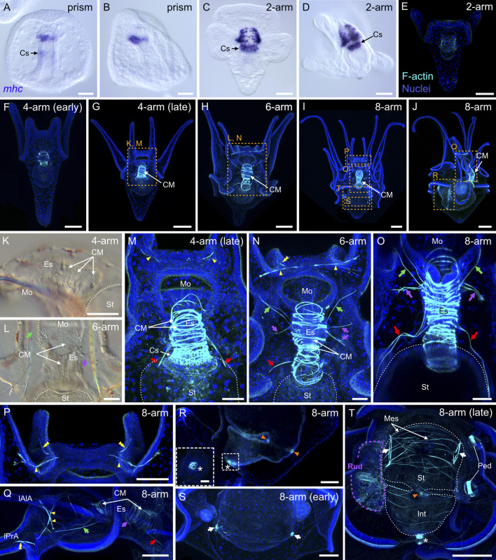
FIGURE 10.
Myogenesis in Paracentrotus lividus during the embryonic and larval periods. Developmental stages are as follows: (A,B) prism stage (prism); (C–E) 2-arm pluteus stage (2-arm); (F,G,K,M) 4-arm pluteus stage (4-arm); (H,L,N) 6-arm pluteus stage (6-arm); (I,J,O–T) 8-arm pluteus stage (8-arm). The use of (early) or (late) associated with the stage names simply highlights here more specific periods during the 4- or 8-arm pluteus stages. In (A–D), images were acquired using bright-field, differential interference contrast light, and they correspond to embryos and larvae labeled for the muscle terminal differentiation gene myosin heavy chain (mhc). In (E–J,M–T), images are maximum intensity projections of confocal z-stacks of larvae co-labeled for F-actin (muscles; cyan) and DNA (nuclei; blue), and they correspond to projections of the entire specimen. In (K,L), images were acquired using bright-field light microscopy. In (A), the embryo is in ventral view, with the animal pole up. In (B,D,J,K,Q,R), embryos and larvae are in left view, with either the animal pole up and the ventral side left in (B) or with the anterior pole up and the ventral side left in (D,K,Q,R) or with the anterior pole right and the ventral side up in (J). In (C,E–I,L–P,S,T), larvae are in anterior view, with the ventral side up. (K–T) Close-ups of the regions outlined by orange boxes in (G–J). (K–O,Q) Close-ups of the esophageal region. (P) Close-up of the oral hood. (R) Close-up of the intestinal region. ((R) inset) Close-up of the anal sphincter. (S,T) Close-ups of the stomach region. In (K–O,T), the white dotted line outlines the larval digestive tract. In (L,N,O,Q), green arrows highlight the posterior dilator muscles, and purple arrows mark the lateral muscles. In (M–O,Q), red arrows indicate the ventrolateral processes (or longitudinal musculature). In (M,N,P,Q), yellow arrowheads mark the anterior dilator muscles (or star-shaped muscles). In (N,P,Q), yellow double arrowheads highlight the preoral dilator muscles. In (R,T), orange arrowheads mark the pyloric sphincter, and the white asterisk marks the anal sphincter. In (S,T), white arrows indicate the lateral stomach muscles. In (T), the purple dotted line delineates the adult rudiment. In (H,J,M,N,P,Q,S,T), the F-actin staining detected along the ciliary band and the epaulettes corresponds to a counterstain of the apex of the cuboidal cells and thus not to muscle cells. Scale bar: (A–D,M–O) 30 μm; (E,F,K,L,P–T) 50 μm; (G–J) 100 μm; ((R) inset) 10 µm. CM: circumesophageal muscle; Cs: cardiac sphincter; Es: esophagus; Int: intestine; lAlA: left anterolateral arm; lPrA: left preoral arm; Mes: mesentery; Mo: mouth; Ped: pedicellariae; Rud: adult rudiment; St: stomach.

