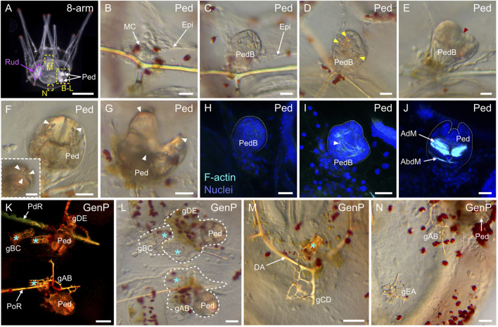
FIGURE 14.
Development of the pedicellariae and the genital plates in Paracentrotus lividus larvae. In (A–N), images are from larvae at the 8-arm pluteus stage (8-arm). In (A), the image was acquired using dark-field light microscopy. In (B–G,L–N), images were obtained using bright-field light microscopy. In (H–J), images are maximum intensity projections of confocal z-stacks of larvae co-labeled for F-actin (muscles; cyan) and DNA (nuclei; blue), and they correspond to projections of the entire pedicellaria. In (K), the image was taken using polarized light to highlight the skeletal elements. In (A), the larva is in anterior view, with the ventral side up and the left side to the left. (B–N) Close-ups of the regions outlined by yellow boxes in (A). (B–J) Close-ups of pedicellariae (Ped). (K–N) Close-ups of genital plates (GenP). In (A), the purple dotted line delineates the adult rudiment on the left side of the larva. In (D), yellow arrowheads indicate the skeletal elements developing inside the pedicellaria bud. In (E), the red arrowhead shows the individualization of the three lobes within the bud. In ((F), (F) inset, (G)), white arrowheads mark the three jaws of the pedicellaria, which can be either closed like in (F) and ((F) inset) or open like in (G). In (H–J), white dotted lines outline the pedicellaria bud in (H–I) or the pedicellaria jaws in (J). In (I), the white arrowhead points to the first muscle fibers appearing within a pedicellaria bud. In (K–M), cyan asterisks indicate the position of juvenile spines associated with the genital plates. In (L), white dotted lines delineate the genital plates and their associated juvenile spines developing in the vicinity of pedicellariae. Scale bar: (A) 200 μm; ((B–G), (F) inset) 15 μm; (H–J,K–M) 30 μm; (N) 50 µm. AdM: adductor muscle; AbdM: abductor muscle; DA: dorsal arch; Epi: larval epidermis; gAB: genital plate AB; gBC: genital plate BC; gCD: genital plate CD; gDE: genital plate DE; gEA: genital plate EA; GenP: genital plate; MC: mesenchyme cell; PdR: posterodorsal rod; Ped: pedicellaria; PedB: pedicellaria bud; PoR: postoral rod; Rud: adult rudiment.

