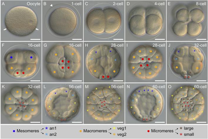
FIGURE 4.
Early cleavage stages of Paracentrotus lividus under light microscopy. Developmental stages are as follows: (A) unfertilized egg (oocyte); (B) zygote (or 1-cell stage) (1-cell); (C) 2-cell stage (2-cell); (D) 4-cell stage (4-cell); (E) 8-cell stage (8-cell); (F,G) 16-cell stage (16-cell); (H,I) 28-cell stage (28-cell); (J,K) 32-cell stage (32-cell); (L,M) 56-cell stage (56-cell); (N,O) 60-cell stage (60-cell). In (A–F,H,J,L,N), the embryos are in lateral view with the animal pole up. In (G,I,K,M,O), the embryos are in vegetal view. In (A), arrows highlight the equatorial pigment band. In (B), the arrowhead marks the fertilization envelope. In (F–K), dots in blue, orange, and red respectively indicate: the mesomeres, the macromeres, and the micromeres. In (H–O), asterisks in dark blue, light blue, yellow, orange, brown, and red respectively mark: the an1, an2, veg1, and veg2 cells as well as the large and the small micromeres. The schematic legend below the images illustrates cell lineage relationships. Scale bar: (A–O) 30 μm. an: animal; veg: vegetal.

