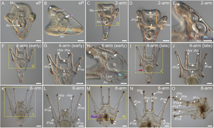
FIGURE 7.
Paracentrotus lividus larval development under light microscopy. Developmental stages are as follows: (A,B) early pluteus stage (eP); (C–E) 2-arm pluteus stage (2-arm); (F–J) 4-arm pluteus stage (4-arm); (K,L) 6-arm pluteus stage (6-arm); (M–O) 8-arm pluteus stage (8-arm). The use of (early) and (late) associated with the stage names simply highlights here more specific periods during the 4-arm pluteus stage. In (A,C,D,F,G,I–N), the larvae are in anterior view with the ventral side up. In (B,E,H,O), the larvae are in left view, with the ventral side left and the anterior side up. In (B,D,E,G,H,J,L,N), the asterisk marks the oral hood located above the mouth. In (B,E,H), the white arrow indicates the cardiac sphincter separating the esophagus and the stomach, and the cyan arrow marks the pyloric sphincter separating the stomach and the intestine. In (B), pink arrowheads highlight red-pigmented cells present within the dorsal ectoderm. In (D,G,J,L,N), the images correspond to close-ups of the regions outlined by yellow boxes in (C,F,I,K,M), respectively. In (E,F,H,I,K,M), the white dotted line outlines the stomach. In (I), the red arrow designates the larval stomach region. In (M), the purple dotted line highlights the adult rudiment, and white arrowheads mark the pedicellariae. Scale bar: (A–E,H) 30 μm; (F,G,I,J) 50 μm; (K–O) 100 µm. An: anus; Es: esophagus; Int: intestine; lAla: left anterolateral arm; lPda: left posterodorsal arm; lPoa: left postoral arm; lPra: left preoral arm; Mo: mouth; pr: postoral region; rAla: right anterolateral arm; rPda: right posterodorsal arm; rPoa: right postoral arm; rPra: right preoral arm; Rud: adult rudiment; St: stomach.

