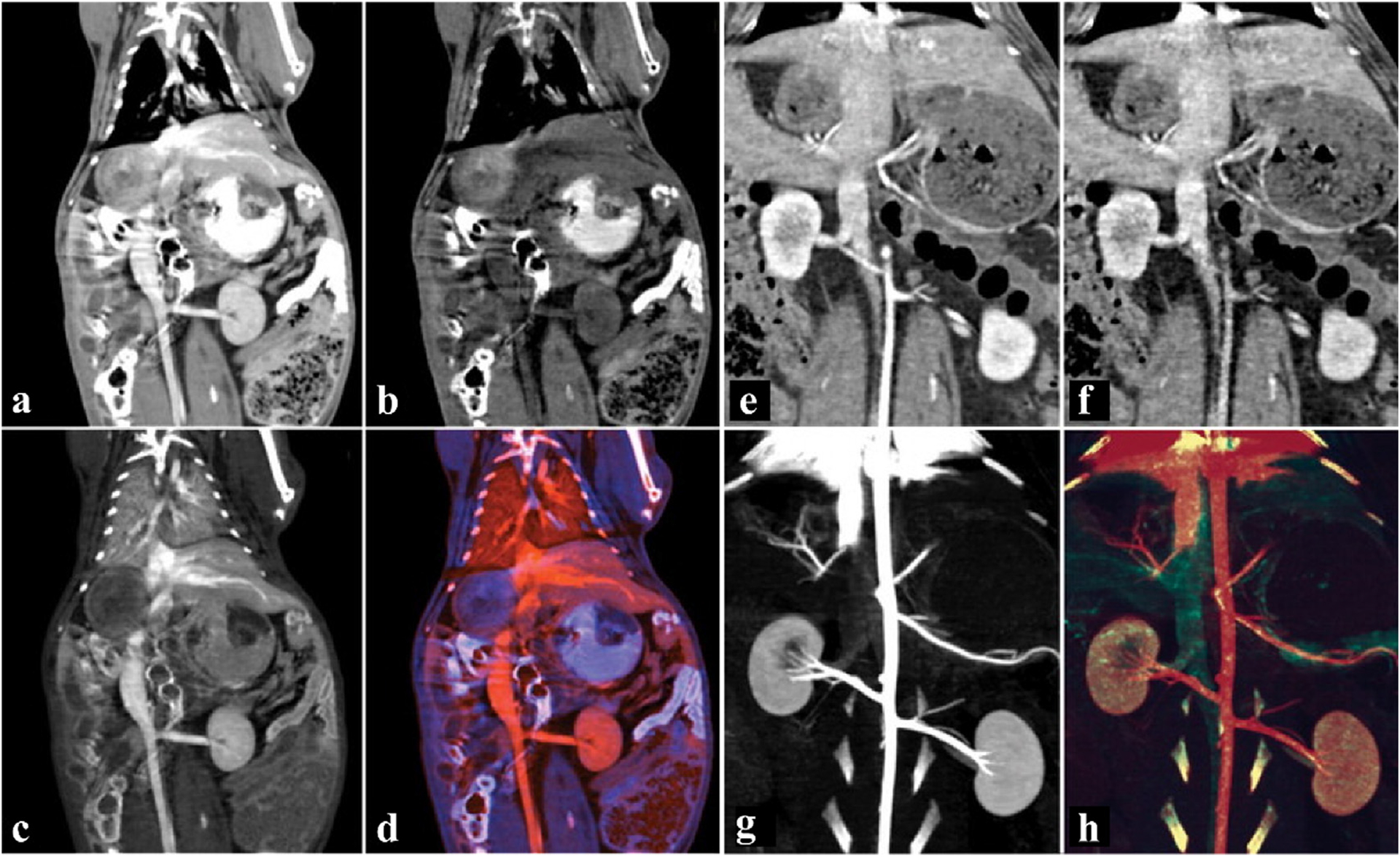
Fig. 14.

Studies on tungsten NPs CAs. a-d) Coronal images of the abdomen with bismuth-based and I-based CAs. a) Virtual monochromatic image at 70 keV. b) Bismuth material map. c) I material map. d) The overlaid color map of bismuth and I. e-h) Angiographic coronal image of the abdomen with W-based and I-based CAs. e) Virtual monochromatic image at 140 keV. f) W material map. g) I material map. h) The overlaid color map of W and I (The red area is iodine, the blue-purple area is bismuth, and the green area is W) (GE Healthcare). Reproduced from Ref. [140] Copyright 2012, Radiology.
