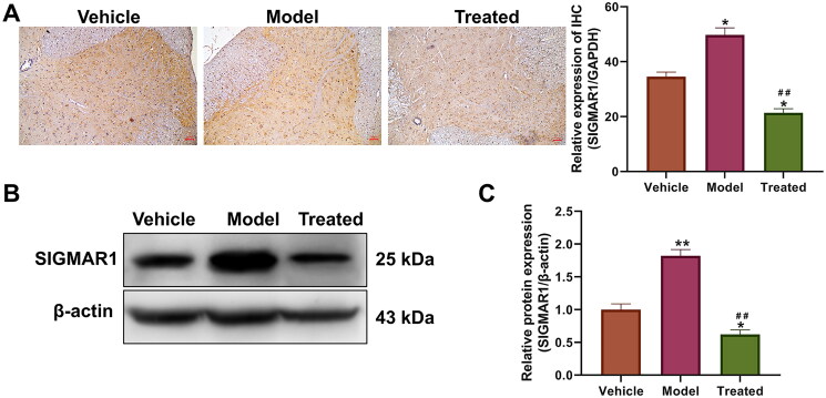
Figure 4.
Effect of veratramine on SIGMAR1 protein expression in spinal cord tissue of diabetic rats. (A) Immunohistochemistry (IHC) was used to stain and localise SIGMAR1 protein in rat spinal cord tissue, and the figure on the right is a statistical quantification figure of IHC results. (B) WB was used to detect the protein expression level of SIGMAR1. (C) Statistical quantification results of grey values of WB bands. **p < 0.01, *p < 0.01, vs. vehicle group; ##p < 0.01, vs. model group. The scale bar is 50 μm.

