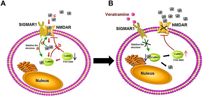
Figure 6.
Mechanism of action of veratramine on the SIGMAR1-NMDAR pathway. A mechanistic diagram of the SIGMAR1-NMDAR pathway in the pathologically induced DPN state. (A) SIGMAR1 can stabilise the intracellular Ca2+-CAMKII structure. (B) The stable Ca2+-CaMKII complex structure leads to decreased CaMKII in the free state. The inhibitory effect of CaMKII protein on NMDAR is weakened, and the activity of NMDAR increases. (C) After NMDAR activity is increased, extracellular Ca2+ flows into the cell. (D) Increased intracellular Ca2+ concentration consolidates the SIGMAR1-NMDAR complex structure and further enhances NMDAR activity. (B) Effects of veratramine treatment on the SIGMAR1-NMDAR pathway: After SIGMAR1 activity is inhibited by veratramine, the stability of the Ca2+-CaMKII complex structure is decreased, and the free CaMKII protein is increased, thus inhibiting NMDAR activity. When NMDAR activity is decreased, Ca2+ inflow into cells decreases, and the stability of the SIGMAR1-NMDAR complex structure decreases, further weakening NMDAR protein activity.

