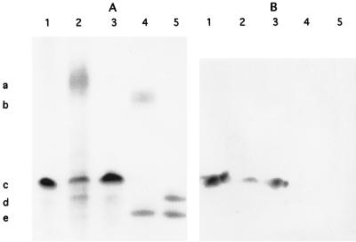
FIG. 2.
(A) SDS-PAGE–silver stain analysis of LPS from B. pertussis Tohama I (lane 1), B. bronchiseptica wild type (lane 2) and mutant (lane 3), and B. parapertussis wild type (lane 4) and mutant (lane 5). The markers indicate the migratory positions of the O antigen of B. bronchiseptica (a, lane 2) and B. parapertussis (b, lane 4), band A of B. pertussis (c, lane 1) and B. bronchiseptica (c, lanes 2 and 3), band B of B. pertussis (d, lane 1) and B. bronchiseptica (d, lanes 2 and 3), the novel structure expressed by the B. parapertussis mutant (d, lane 5), and the truncated band B of B. parapertussis (e, lanes 4 and 5). Wild-type B. bronchiseptica and B. parapertussis both expressed O antigen whereas the mutants did not. (B) Western blot analysis of a replica of the gel shown in panel A with monoclonal antibody BL-2, which recognizes an epitope in band A. This analysis demonstrates that the B. bronchiseptica O-antigen mutant was not affected in band A expression and that the novel structure expressed by the B. parapertussis O antigen mutant was not a full band A structure.

