
Figure 5. Microtubule detyrosination inhibits katanin-mediated severing and polyglutamylation can overcome this inhibition.
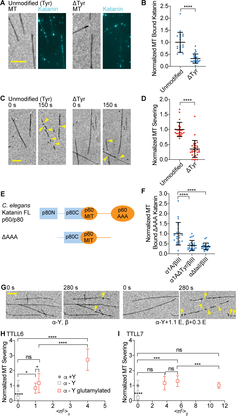
(A) Katanin association to unmodified tyrosinated and detyrosinated microtubules. Scale bar, 5 μm.
(B) Normalized katanin bound to tyrosinated and detyrosinated microtubules. Assays at 4 nM katanin, 1 mM ATP; n = 19 tyrosinated, 24 detyrosinated microtubules from multiple chambers.
(C) Severing of unmodified tyrosinated and detyrosinated microtubules. Yellow arrows, severing. Scale bar, 5 μm.
(D) Normalized severing of tyrosinated and detyrosinated microtubules. Reactions at 20 nM katanin, 1 mM ATP; n = 24, 31 tyrosinated and detyrosinated microtubules, respectively, from multiple chambers. LC-MS of microtubules and additional data in Figure S6.
(E) Domain organization of C. elegans katanin showing interactions between p60 and p80 subunits.
(F) Microtubule binding of p60ΔAAA/p80Cterm to recombinant human tyrosinated (α1A/βIII), detyrosinated (α1AΔY/βIII), and microtubules missing their α-tubulin tails (α1AΔtail/βIII); n = 22, 24 and 22 tyrosinated, detyrosinated and α-tailless microtubules, respectively from multiple chambers.
(G) Severing of detyrosinated microtubules (left) and detyrosinated microtubules glutamylated by TTLL6 (right). Yellow arrows, severing. Scale bar, 5 μm.
(H, I) Severing of detyrosinated microtubules with progressively higher glutamylation levels added by TTLL6 (H) or TTLL7 (I), normalized to severing of unmodified microtubules. Filled circle, tyrosinated, open square, detyrosinated microtubules with different glutamylation levels; Severing at 20 nM katanin, 1 mM ATP; n ≥ 29 microtubules from multiple chambers. All error bars, S.D. p-value > 0.05 (ns), p ≤ 0.05 (*), 0.001 (***), 0.0001 (****) by Mann-Whitney test.