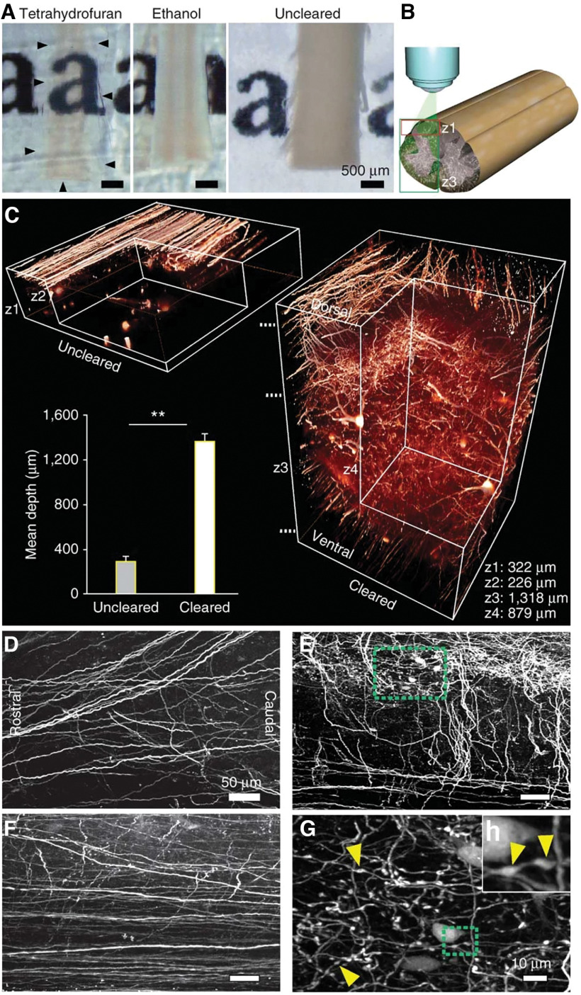
Figure 1.
Clearance allows high-resolution imaging of the unsectioned spinal cord. A, Clearance with THF renders the adult mouse spinal cord transparent (outlined by black arrowheads) while ethanol-treated tissue remains opaque (photographic images are shown). B, Schematic illustration of the two-photon–imaged regions and depths: only dorsal (z1) or entire dorsoventral (z3) spinal cord. C, Comparison between uncleared (left) and cleared (right) spinal cords of GFP-M mice imaged with two-photon microscopy. Values are mean ± SD. **p < 0.01. D–G, Horizontal projections from the cleared spinal cord at different depths marked in C: dorsal (∼100 μm; D), mid (∼500 μm; E), and ventral (∼1200 μm; F). G, Higher magnification of the area marked in green in E. H, Higher magnification of the area marked in green in G. Yellow arrowheads in G, H depict some of the axonal boutons in the gray matter. From Ertürk et al. (2011).

