Figure 4.
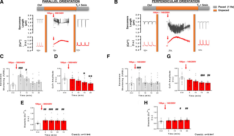
Effect of high-voltage pulsed electric field on sarcomere shortening and intracellular Ca2+ ([Ca2+]i) in left ventricular myocytes according to cell orientation. Representative sarcomere lengths and [Ca2+]i traces obtained from an isolated left ventricular myocytes (LVM) during baseline (control [Ctrl]) 1 Hz pacing, during and after (T0+5min) a 100 µs high voltage electric pulse (EP) delivered to a parallel (A) and perpendicular-oriented cell (B) below their respective lethal threshold. Sarcomere shortening, expressed as a percentage of resting sarcomere length (%RSL) was increased 5 minutes after EP and returned to Ctrl level 15 minutes post-EP in parallel-oriented cells (C). Ca2+ transients (CaTr) amplitude progressively decreased after EP (D) while diastolic levels increased (E). Similar trends on sarcomere shortening (F), CaTr amplitudes (G) and diastolic Ca2+ levels (H) were observed in perpendicular-oriented cells. Data are represented as mean±SE of the mean with individual values for each cell. Repeated-measure 1-way ANOVA: *P<0.05, **P<0.01, #P<0.05, ##P<0.01, ###P<0.001. N indicates number of animals; n, number of cells; and r.u., ratio units.

