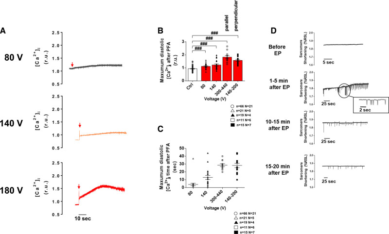
Figure 5.
Immediate effect of a monophasic single electric pulse (EP) on left ventricular myocyte diastolic Ca2+ levels and spontaneous contractile activity. A, Representative intracellular Ca2+ ([Ca2+]i) traces from left ventricular myocytes (LVMs) subjected to a low- (80 V), medium- (140 V), and high-voltage (here, 180 V in a perpendicular cell) 100 µs EP. B, The rise in diastolic Ca2+ level induced by the EP increased when increasing pulse’s amplitude. C, The time to reach maximum diastolic Ca2+ level also tended to increase with the voltage of the electroporating pulse. D, Example of spontaneous contractions monitored before and after EP (intermediate voltage, 100 µs) in unpaced LVM. Prior to EP, when pacing was stopped, LV myocytes remained quiescent. Spontaneous contractions were observed in these cells 1–5 minutes after EP and their frequency decreased with time over 20 minutes. Data are represented as mean±SE of the mean with individual values for each cell. Unpaired t test: ###P<0.001. N indicates number of animals; n, number of cells; PFA, pulsed field ablation; and r.u., ratio units.

