
Fig. 3.
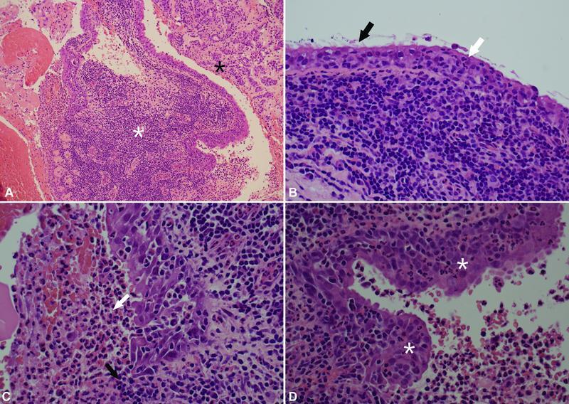
Histology of the specimen. ( A ) Histology illustrates a benign cyst with acute-on-chronic inflammation ( white asterisk ) in the vicinity of atrophic/fibrotic anterior pituitary gland tissue ( black asterisk ). ( B ) The cyst epithelium is focally ciliated ( black arrow ) and acutely inflamed by infiltrating neutrophils ( white arrow ). Underneath, there is a dense chronic inflammatory infiltrate. ( C ) Focally, the epithelium is eroded by inflammation ( black arrow ), and purulent material is seen in the cyst lumen ( white arrow ). ( D ) The epithelium shows squamous metaplasia and mild reactive atypia in places ( white asterisk ), as secondary changes to the inflammation.