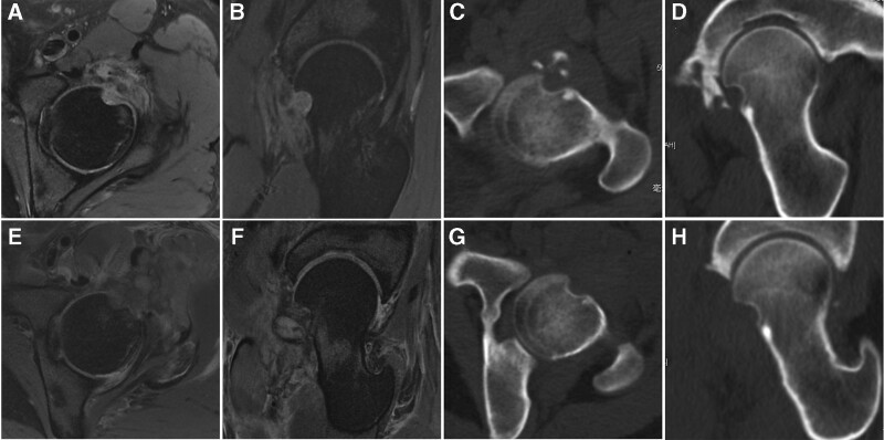
Figure 1.
(A, B) Preoperative axial and oblique sagittal MRI showed soft tissue mass in the femoral head neck junction. (C, D) Preoperative axial and oblique sagittal CT showed adjacent bone defect and ossification. (E, F) Postoperative axial and oblique sagittal MRI showed total excision of the mass. (G, H) Postoperative axial and oblique sagittal CT showed total excision of the mass. CT = computed tomography, MRI = magnetic resonance images.

