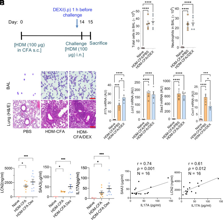
FIGURE 3.
The expressions of Lcn2 and Saa3 were resistant to steroid treatment in the preclinical type 17 severe asthma model. (A–C) Eight-week-old female WT C57BL/6 female mice were subjected to the HDM-CFA acute asthma model. PBS or DEX was administered to the mice (as described in Materials and Methods). Twenty-four hours after the challenge, total cell and neutrophil counts in the BAL were quantified (B), representative BAL cells were prepared by cytospin, and lung tissues were subjected to histochemical staining as indicated (C). All scale bars (red), 100 μm. (D) mRNA expression of lung tissues was quantified by RT-PCR. (E) Plasma LCN2, SAA3, and IL-17A levels were measured using ELISA. One-way ANOVA was performed, followed by Tukey’s multiple-comparisons test or Kruskal–Wallis test (nonparametric) followed by Dunn’s multiple-comparisons test. (F) The correlation between plasma LCN2 or SAA3 and IL-17A was performed using the Pearson correlation test. For (B), (D) and (E), *p < 0.05, **p < 0.01, ***p < 0.001, ****p < 0.0001. AU, fold induction relative to unchallenged control mice.

