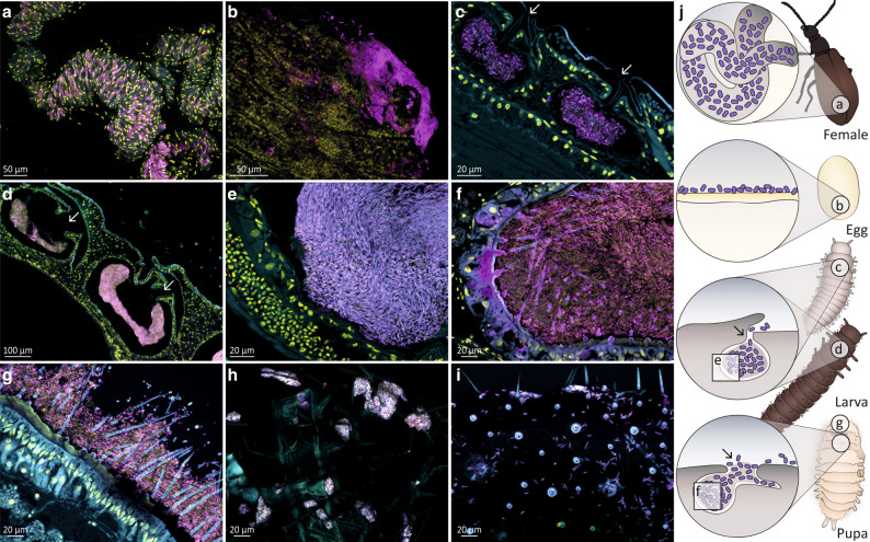
Fig. 4. Burkholderia Lv-StB symbiont localization and prevalence throughout the different life stages of L. villosa using fluorescence in situ hybridization.
Symbiont cells are generally depicted in magenta, and host cell nuclei in yellow. a Whole mount of a female abdomen showing a dense population of Lv-StB in tubes of the accessory glands. b Whole-mount of an egg revealing Lv-StB dominance. c Sagittal section of a 1st instar larva revealing a dense culture of Lv-StB in the dorsal symbiotic structures as well as an opening to the external environment (white arrows). d Sagittal sections through the three pouches of a later larval stage showing the same morphology and (e) the dominance of Lv-StB inside the pouch. f 1st dorsal symbiont compartment and (g) outer surface of a pupa showing dominance of Lv-StB. In (a–g) B. gladioli specific staining is shown in cyan (Burk16S_Cy3), Lv-StB-specific staining in magenta (Burk16S_StB_2_Cy5), and host cell nuclei in yellow (DAPI). h First larval exuvia covered with B. gladioli. i Inner side of a larva-pupa exuvia with visible symbionts cells. FISH on exuviae (h, i) show general eubacterial staining in cyan (EUB338_Cy3) and B. gladioli specific staining in magenta (Burk16S_Cy5). j Schematic guide illustrating symbiont localization throughout L. villosa development. 1st instar larva individuals are from the 1st lab generation, and all other individuals are field collected. In every image autofluorescence of the host tissue is shown in cyan and overlap of all three channels is shown in purple-white. Arrows indicate the opening of the symbiotic structures in larvae and pupae. Scale bars correspond to 20 µm, except for panels a (50 µm) and (d) (100 µm).

