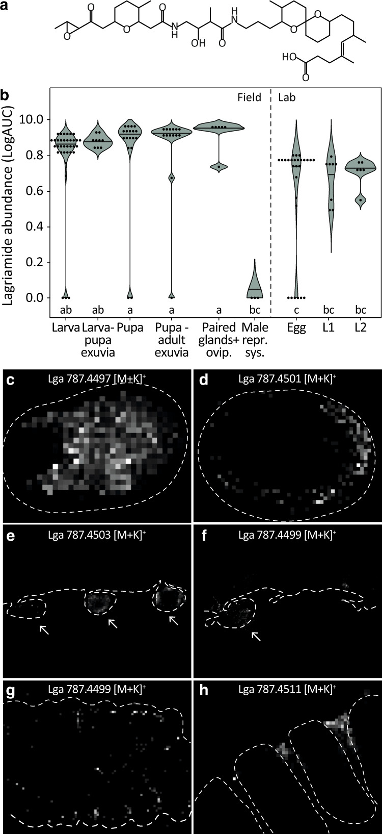
Fig. 5. Lagriamide is present across Lagria villosa life stages and co-localizes with Lv-StB in regions exposed to the external environment.

a Chemical structure of lagriamide [11]. b Area under curve (AUC) of the extracted ion chromatogram (EIC) of lagriamide (m/z = 747.4769–747.4843 [M-H]−) representing abundance across host development (Table S6), as quantified from crude methanol extracts. Eggs, as well as first (L1) and second (L2) instar larvae correspond to offspring from field-collected females. All others correspond to field specimens. Different letters indicate significant differences between life stages (Kruskal-Wallis χ2 = 66.988, df = 8, p value = 1.95e–11, posthoc Dunn’s Test, α ≤ 0.05). c–h 2-D ion maps obtained by AP-SMALDI-MSI representing the potassium adducts of lagriamide [M + K]+ across L. villosa life stages. c Surface analysis of an intact egg and (d) an egg cryosection showing lagriamide presence on the surface. e In larval sections, lagriamide is present inside the symbiotic organs (arrows). f In pupal sections, lagriamide is mainly detected in the first symbiotic organ (arrow). g On the inner surface of a larva-to-larva exuvia, lagriamide is either scattered over the thoracal segments or (h) distinctly located between the thoracic segments and first abdominal segment, which corresponds to the location of the symbiotic organs. Dotted lines were manually added based on corresponding light microscopy pictures (Fig. S6) to indicate specimen profiles.
