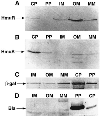
FIG. 5.
Localization of HmuR and HmuS in Y. pestis cellular fractions by Western blot analysis. Iron-depleted Y. pestis KIM6+(pRT240) cells were grown in deferrated PMH broth at 30°C. Periplasm (PP), cytoplasm (CP), IM, OM, and mixed-membrane (MM) fractions were isolated, and equal concentrations of proteins in each cell fraction were separated in an SDS–12% polyacrylamide gel. The immunoblot was reacted with anti-HmuR antiserum (A), anti-HmuS antiserum (B), anti-β-galactosidase (β-gal) IgG1 antibody (C), or anti-β-lactamase (Bla) polyclonal antibody (D).

