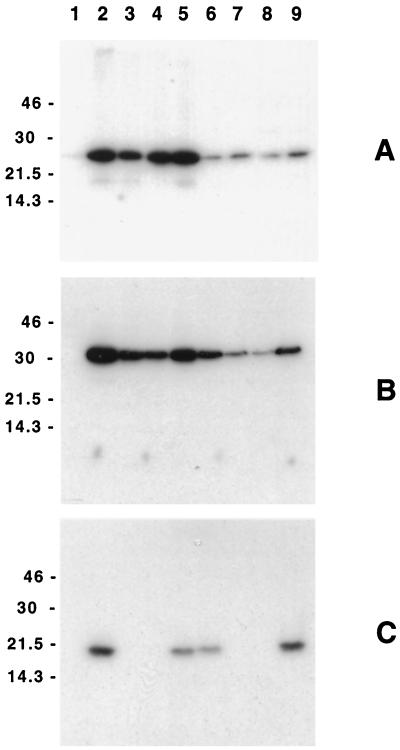
FIG. 2.
Western blot analysis of the H. ducreyi CdtABC proteins expressed by selected E. coli and H. ducreyi strains. Whole-cell lysates of these strains were probed with the H. ducreyi CdtA-reactive MAb 1G8 (A), the H. ducreyi CdtB-reactive MAb 20B2 (B), and the H. ducreyi CdtC-reactive MAb 8C9 (C). Lanes: 1, E. coli DH5α(pBR322); 2, E. coli DH5α(pJL300); 3, E. coli DH5α(pJL303); 4, E. coli DH5α(pJL303)(pLS88); 5, E. coli DH5α(pJL303)(pJL300-C); 6, wild-type H. ducreyi 35000; 7, H. ducreyi cdtC mutant 35000.303; 8, H. ducreyi 35000.303(pLS88); 9, H. ducreyi 35000.303(pJL300-C). Molecular size markers (in kilodaltons) are listed on the left of each panel.

