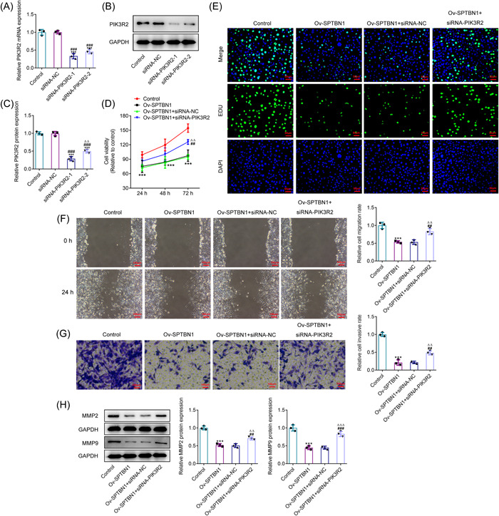
Figure 5.

SPTBN1 attenuated the proliferation, invasion, and migration of RA‐FLSs via PIK3R2. (A–C) The expression of SPTBN1 in RA‐FLSs transfected with siRNA‐PIK3R2‐1 or 2 was detected using RT‐qPCR and western blot. ***p < .001 versus Control group. ### p < .001 versus siRNA‐NC group. (D) The viability of RA‐FLSs transfected with Ov‐SPTBN1 and siRNA‐PIK3R2 was detected using CCK‐8. (E) The proliferation of RA‐FLSs transfected with Ov‐SPTBN1 and siRNA‐PIK3R2 was detected using Edu (magnification, 200×). (F, G) The migration and invasion of RA‐FLSs transfected with Ov‐SPTBN1 and siRNA‐PIK3R2 were detected using wound healing and transwell (magnification, 100×). (H) The expressions of MMP2 and MMP9 in RA‐FLSs transfected with Ov‐SPTBN1 and siRNA‐PIK3R2 were detected using western blot. ***p < .001 versus Control group. ## p < .01 and ### p < .001 versus Ov‐SPTBN1 group. ∆∆ p < .01 and ∆∆∆ p < .001 versus Ov‐SPTBN1 + siRNA‐NC group. n = 3. ANOVA followed by Tukey's test. ANOVA, analysis of variance; CCK‐8, Cell Counting Kit‐8; FLS, fibroblast‐like synoviocyte; RA, rheumatoid arthritis; RT‐qPCR, reverse transcription‐quantitative PCR.
