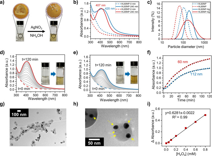
Figure 4.
Self-assembly of silver nanoparticles on the surface of cross-linked HLNPs. (a) Scheme of formation of silver nanoparticles on the surface of LNPs. (b) UV–vis spectra of HLNPs before and after silver nanoparticles are assembled on the particle surface. (c) DLS particle size analysis of HLNPs before and after self-assembly of silver nanoparticles on the surface. (d) UV–vis spectra of silver nanoparticles formed on the HL85NP surface with a 60 nm diameter (data shown at 10 min intervals). (e) UV–vis spectra of silver nanoparticles formed on the HL85NP surface with a 110 nm diameter (data shown at 10 min intervals). (f) UV–vis kinetic plot of silver nanoparticle self-assembly on HL85NP with different particle sizes. (g) TEM image of HLNPs reacted with silver ammonia solution. (h) Silver nanoparticles formed on the surface of HLNPs with yellow arrows pointing on them. (i) Calibration curve for the detection of H2O2 using HLNP–silver hybrid nanoparticles.

