
Fig. 2.
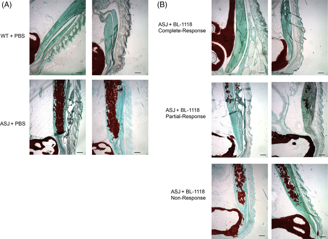
(A) Histopathology of representative Achilles entheses in 23-week-old WT (top) and Enpp1asj/asj mice (bottom) treated with vehicle. Calcific deposits are highlighted by Alizarin Red deposits embedded in the Achilles tendon, which appears green. (B) Histopathology of representative Achilles entheses in 23-week-old Enpp1asj/asj mice treated with weekly subcutaneous injections of 0.3 mg/kg BL-1118. Top panels illustrate representative histologic response in complete responders, middle panels illustrate the histologic response in partial responders, and bottom panels illustrate the histologic response in nonresponders. Scale bar = 10 μm.