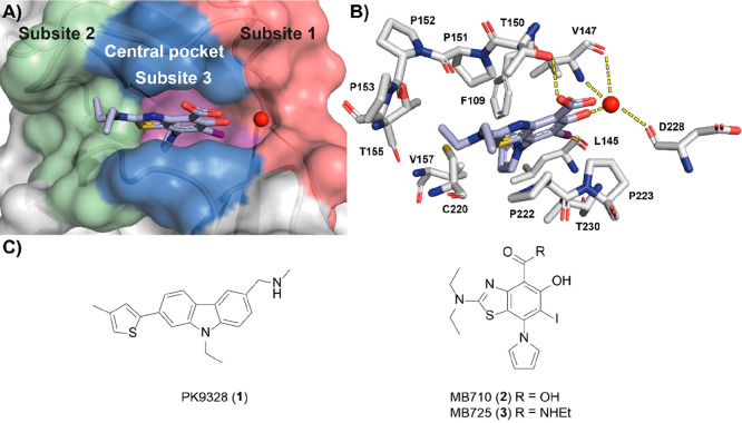
Figure 1.
(A) Crystal structure of 2 (blue sticks) in complex with p53-Y220C (PDB: 5O1I, surface representation)42 with subsites referred to throughout the text color-coded: subsite 1 = red, subsite 2 = green, buried subsite 3 = purple, central pocket = blue. A structural water molecule interacting with 2 is shown as a red sphere. (B) Zoom-in crystal structure of 2 (blue sticks) in complex with p53-Y220C with interacting residues shown (PDB: 5O1I, gray sticks).42 The halogen bond to L145 (dashed orange line) and the hydrogen bond network between the salicylate moiety and the protein are shown (dashed yellow lines). (C) Chemical structures of representative iodophenol and carbazole lead molecules 1–3.

