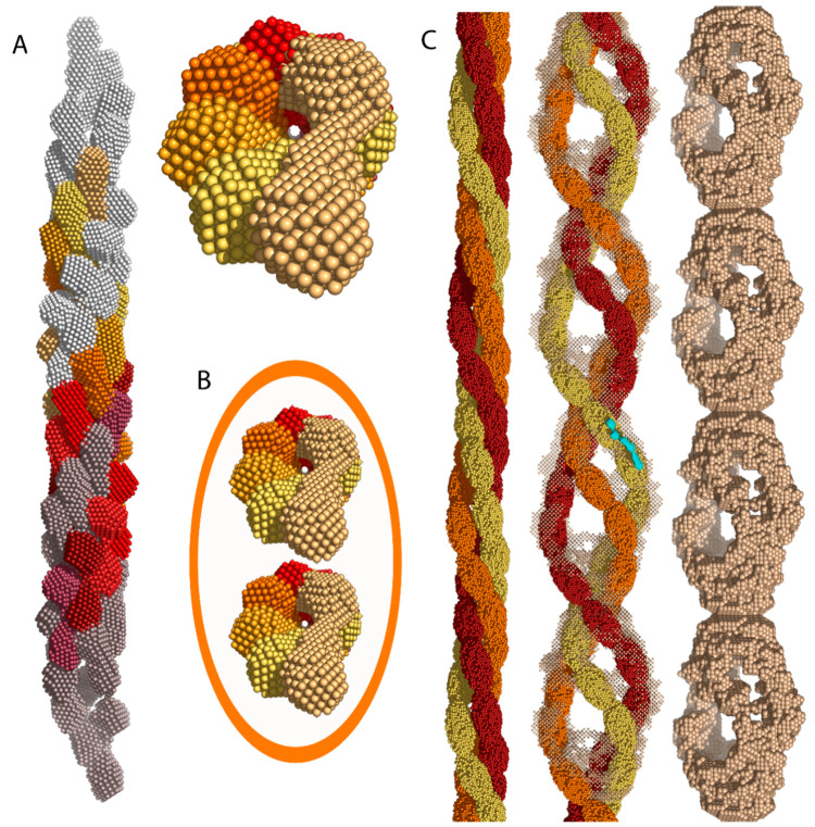
Figure 5.
Successive stages of development in insulin oligomers: protofilaments, protofibrils, and fibrils. (A) Side and top views of an intertwined conglomeration of eight helical insulin oligomers (color scale: purple → red → light yellow) that form a protofilament. The gray segments at the open ends mark additional precursors. (B) Two intertwined protofilaments form a protofibril of 100 Å diameter. The orange ellipse shows the boundaries of an assembled protofibril. (C) Three protofibrils (orange, red, and yellow) interweave to form a mature insulin amyloid fibril. Both the side and frontal views are shown here. Reproduced from ref (62) under an open access Creative Commons License.

