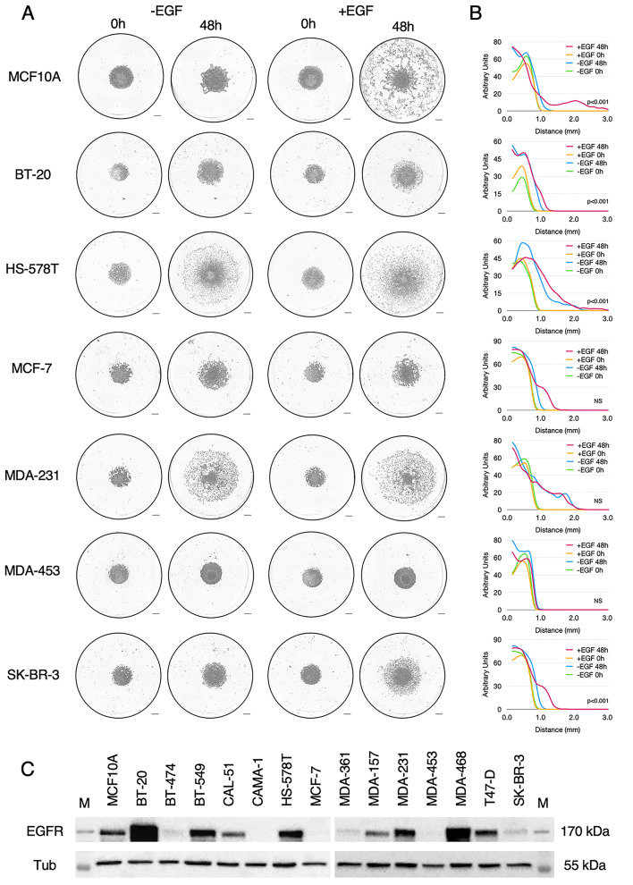
Fig. 7.
Migration of breast cancer cell lines in the aerotactic assay. (A) Representative images taken at 48 h of breast cancer cell lines subjected to the aerotactic assay in breast cancer medium (-EGF) or breast cancer medium supplemented with 10 µg/mL EGF (+ EGF). Scale bars, 0.5 mm. (B) Cell distribution from the centre of the cell cluster (in mm) at 0 h (green) and 48 h (carmine) in the -EGF condition and at 0 h (yellow) and 48 h (blue) in the + EGF condition is indicated as graphs (mean of three experiments). Y-axis scale is expressed in arbitrary unit. To assess the difference between the plus and minus EGF conditions at 48 h, a Student t-test was performed on D5% values. P-values are indicated within graphs. NS non-significant. (C) Western blot analysis of EGFR expression in breast cancer cell lines. Tubulin expression was used as a loading control. M stands for Molecular weight (PageRuler, Thermo). See also Figure S7 for other cell lines

