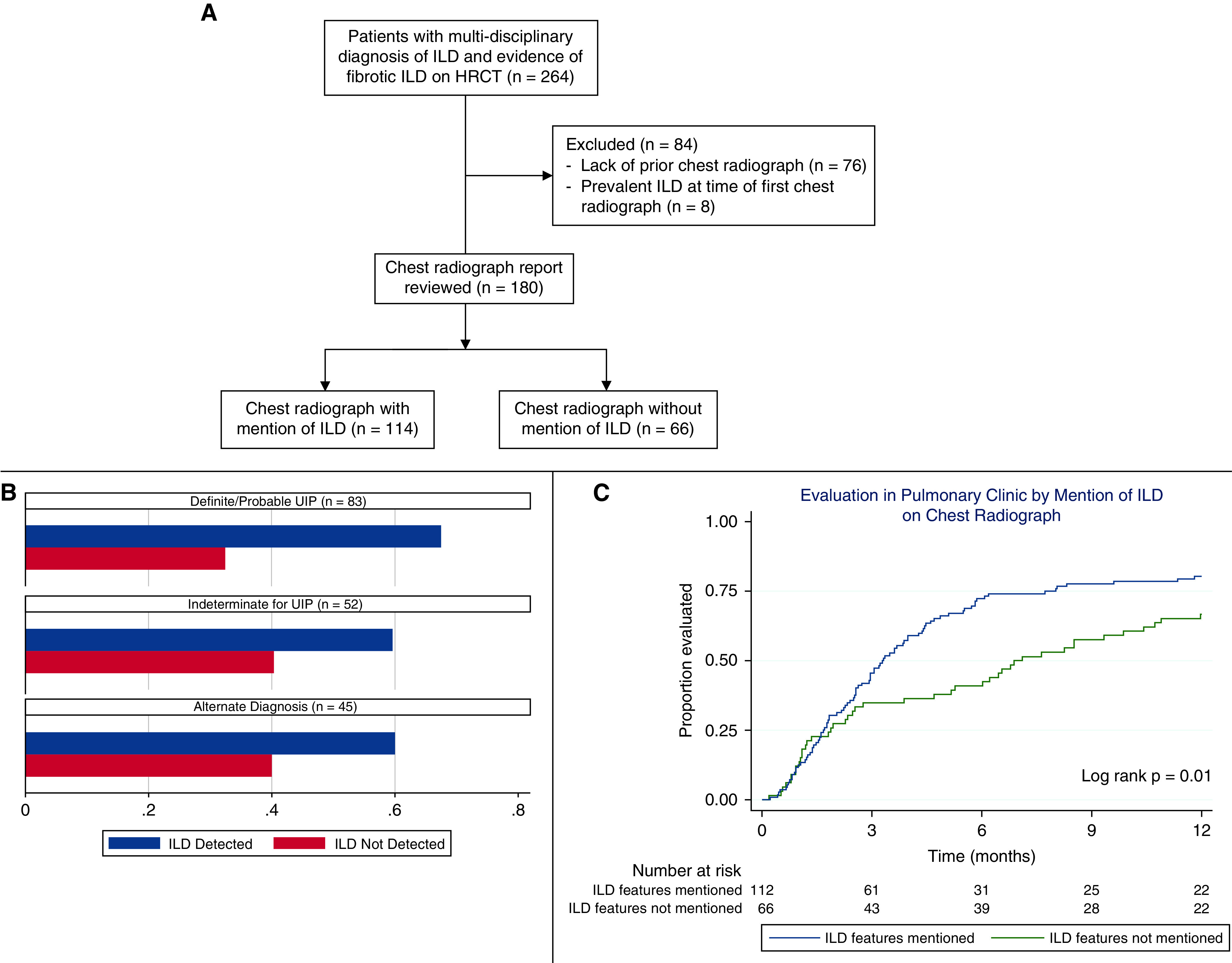
Figure 1.

(A) Flow diagram of eligible and included interstitial lung disease (ILD) cases. (B) Percentage of cases with ILD mentioned on chest radiography, stratified by high-resolution computed tomography pattern. (C) Time to pulmonary evaluation stratified by the mention of ILD on chest radiography. HRCT = high-resolution computed tomography; UIP = usual interstitial pneumonia.
