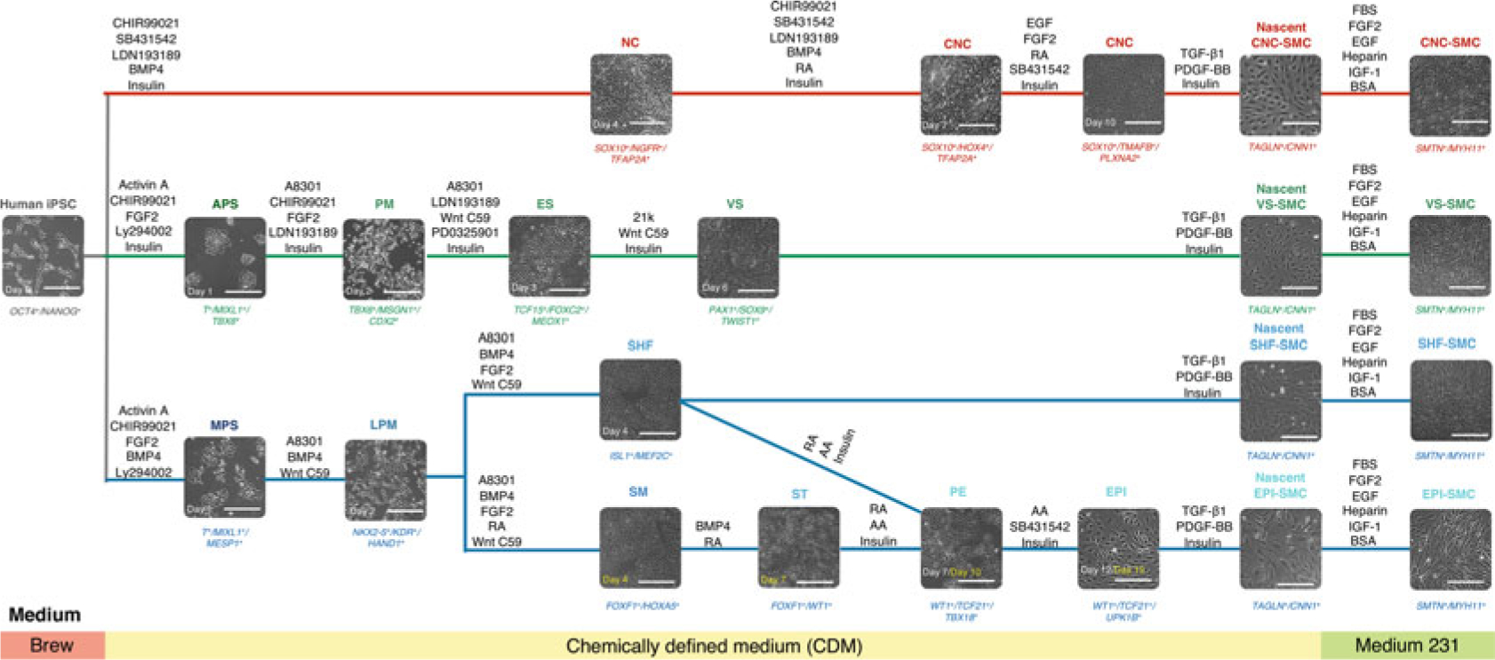
Fig. 1.

Schematic diagram of the conditions for deriving embryonic origin-specific VSMCs from human iPSCs. Lineage-specific intermediate cell types that give rise to each VSMC subtype are differentiated in a stepwise fashion. Each intermediate cell population is subjected to TGF-β1 and PDGF-BB treatment for 6 days before switching to a smooth muscle cell growth medium (Medium 231) for another 14–21 days. Phenotypic markers for specific cell types are represented in italics at the bottom of each bright-field image. NC neural crest, CNC cardiac neural crest, APS anterior primitive streak, PM paraxial mesoderm, ES early somite, VS ventral somite, MPS mid-primitive streak, LPM lateral plate mesoderm, SHF second heart field, SM splanchnic mesoderm, ST septum transversum, PE proepicardium, EPI epicardium. Scale bars represent 50 μm
